
Surgical Lump Removals in Cats and Dogs
Surgical Lump Removal starting from $1,250 + histopathology
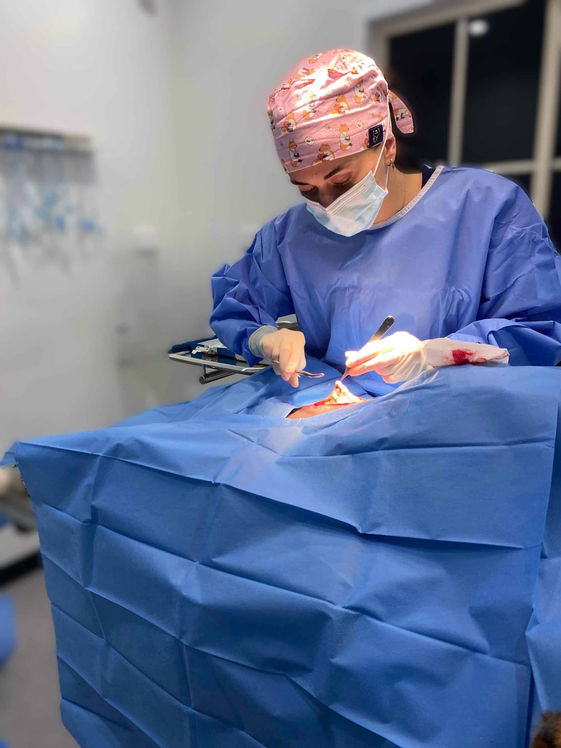
Finding a lump or bump on your pet can be concerning. While not all lumps are dangerous, it's important to have them properly assessed by a veterinarian. At Moreton Bay Vets, we offer surgical lump removal for cats and dogs to diagnose, treat, or prevent potential health problems.
A lump may be a benign (non-cancerous) growth such as a lipoma (fatty tumor), cyst, or wart—but it could also be a sign of a more serious condition like a malignant tumor. The goal of surgical removal is to eliminate the growth, relieve discomfort, and—most importantly—gain a clear diagnosis through lab testing if needed.
Understanding Benign Lumps in Cats and Dogs
Not all lumps are dangerous—in fact, many are benign, meaning they are non-cancerous and typically do not spread to other parts of the body. However, even benign lumps should be assessed by a veterinarian, as they can still cause problems depending on their size, location, or rate of growth.
Some of the most common benign lumps we see in cats and dogs include:
-
Lipomas: These are soft, fatty tumors often found under the skin in middle-aged to older dogs. They tend to grow slowly and are generally harmless, but large lipomas can interfere with mobility or become irritated.
-
Sebaceous Cysts: These occur when a hair follicle or oil gland becomes blocked. They often appear as small, firm bumps and may rupture or become infected if not treated.
-
Warts (Papillomas): Caused by a virus, warts are typically seen in younger animals and can appear around the mouth, face, or paws. They are usually harmless but can be bothersome or multiply quickly.
-
Histiocytomas: Common in younger dogs, these small, raised lumps often appear suddenly and may shrink on their own over time. They are usually benign but can resemble more serious tumors.
Even though benign lumps don’t metastasize (spread), they may still require removal if they:
-
Grow quickly or become very large
-
Are located where they interfere with your pet’s movement or comfort (e.g., under a limb or near a joint)
-
Become ulcerated, infected, or painful
-
Are cosmetically concerning for the owner
-
Cannot be definitively identified without removal and histopathology
In some cases, a benign lump may look similar to a malignant one based on physical exam alone. That’s why we often recommend sampling the lump (via fine-needle aspiration) or sending it for histopathology after removal to be certain of the diagnosis.
Understanding Malignant Tumors in Cats and Dogs
Unlike benign lumps, malignant tumors are cancerous growths that can invade surrounding tissues and, in many cases, spread (metastasize) to other parts of the body, such as the lungs, liver, or lymph nodes. These types of tumors often require more aggressive treatment and close monitoring.
Malignant tumors can vary widely in appearance, location, and behavior, which is why a physical exam alone is not enough to determine if a lump is cancerous. Veterinary surgeons and oncologists rely on tools like biopsy, histopathology, blood work, and imaging to diagnose and stage cancer accurately.
Common Types of Malignant Tumors in Pets
-
Mast Cell Tumors (MCTs): These are one of the most common skin cancers in dogs. They can appear as small, raised bumps or large, ulcerated masses. MCTs vary in aggressiveness and may require surgery, chemotherapy, or radiation depending on the grade.
-
Soft Tissue Sarcomas: These tumors develop in the connective tissues and muscles. They can be deceptively slow-growing but have the potential to invade surrounding tissues. Wide surgical margins are typically needed to reduce the chance of recurrence.
-
Melanomas: While some melanomas (like those on the skin) may be benign, oral or digital melanomas are often malignant and can metastasize quickly, especially in dogs.
-
Squamous Cell Carcinoma (SCC): A locally invasive cancer found in the mouth, skin, or nails. In cats, SCC commonly appears on the ears or nose, especially in light-colored cats exposed to sun.
-
Lymphoma: This cancer affects the lymphatic system and is more commonly found in dogs and cats as a systemic illness rather than a lump. However, it can present as enlarged lymph nodes or skin lesions.
Signs That a Lump May Be Malignant
While not definitive, a lump may be more likely to be malignant if it:
-
Grows rapidly over a short period
-
Is firm, fixed, or attached to underlying tissue
-
Ulcerates or bleeds without healing
-
Changes shape or becomes irregular
-
Causes discomfort or disrupts normal movement or function
The Role of Surgical Removal in Malignant Tumors
If a tumor is suspected or confirmed to be malignant, surgical removal is often the first step. The goal is to excise the mass completely, with clear margins to reduce the risk of recurrence. In some cases, additional treatments like chemotherapy or radiation may be recommended, especially if the tumor is aggressive or has already spread.
Histopathology is particularly crucial when dealing with malignant tumors, as it allows us to identify the exact type, grade, and behavior of the cancer. This helps us determine:
-
Whether the entire tumor was successfully removed
-
The likelihood of recurrence or metastasis
-
Whether further treatment is needed
-
The long-term prognosis for your pet
Early diagnosis and treatment of malignant tumors significantly improve the outcome and quality of life for your pet. If you notice any unusual lumps, changes in existing ones, or other concerning symptoms, we encourage you to schedule an examination as soon as possible.
The Benefits of Lump Removals
-
Early Detection and Treatment: Removing a lump early, especially before it grows or spreads, gives your pet the best chance of a full recovery if the lump turns out to be malignant.
-
Comfort and Mobility: Some lumps can become irritated, infected, or interfere with your pet’s movement. Removal can improve your pet’s comfort and quality of life.
-
Peace of Mind: Knowing whether a lump is benign or malignant helps guide further care decisions and gives you peace of mind.
Before surgery, we can sample the lump through fine-needle aspiration (FNA) to get a preliminary idea of the lump's nature. We also have in-house blood testing machines, allowing us to perform pre-anesthetic blood work quickly and efficiently to ensure your pet is healthy enough for surgery.
What is Histopathology?
Histopathology is the microscopic examination of tissue samples by a veterinary pathologist. After a lump is surgically removed, we can send it to a specialized laboratory where experts analyze the tissue to determine exactly what kind of growth it is.
This process provides a definitive diagnosis, identifying whether the lump is benign or malignant, what type of cells are involved, and whether the growth was completely removed or if further treatment may be needed.
Our team of Veterinarians strongly recommend histopathology for any removed lump because it is the only way to be certain about what the lump was. Even benign-looking lumps can occasionally surprise us with unexpected findings under the microscope. This vital information allows us to tailor ongoing care or monitoring specific to your pet’s needs.
If you've noticed a lump on your pet or have concerns about an existing growth, please contact us to schedule an exam. Early assessment and treatment can make all the difference for your pet’s long-term health.
We accept